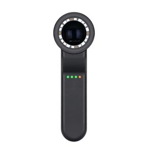
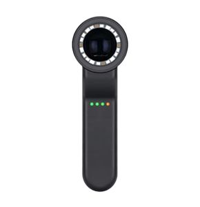
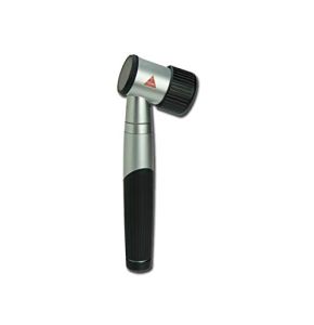
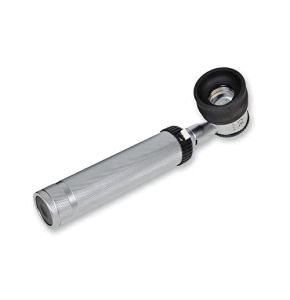
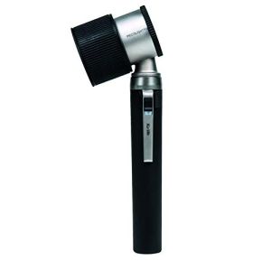
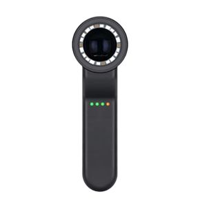
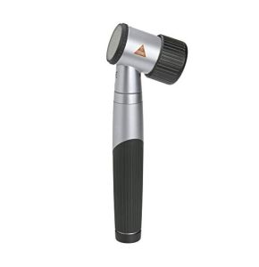
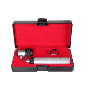
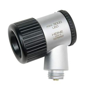
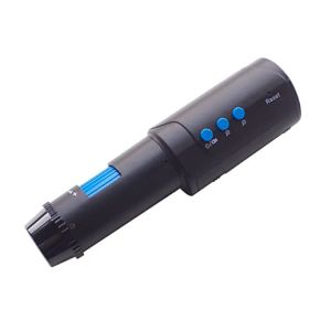
Dermatoscope
18/11/2024 9
18/11/2024 7
18/11/2024 13
18/11/2024 13
18/11/2024 9
18/11/2024 9
18/11/2024 12
18/11/2024 13
18/11/2024 13
18/11/2024 12
18/11/2024 14
18/11/2024 10
18/11/2024 13
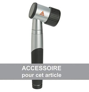
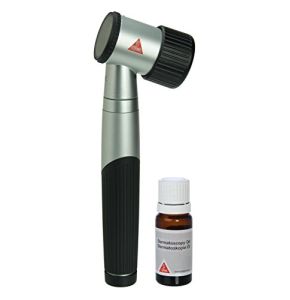
Mini 3000 LED Dermatoscope
18/11/2024 11
18/11/2024 11
18/11/2024 13
18/11/2024 9
Dermatoscope: The Indispensable Device of the Modern Dermatologist
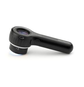
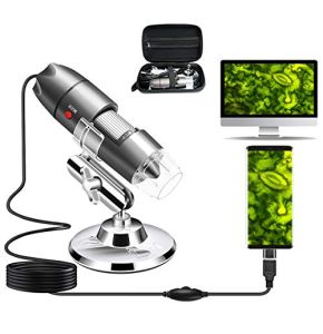
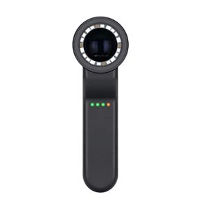
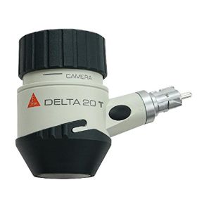
At our dedicated dermatoscope booth, we offer a selection of high-quality devices designed for dermatologists and skin practitioners. Our dermatoscopes combine advanced technology with ease of use, helping you provide precise and rapid diagnoses to your patients. Discover how this essential tool can transform your daily practice.
Why is Dermoscopy Indispensable in Dermatology?
Dermoscopy is a non-invasive examination technique that allows detailed observation of skin structures. With a dermatoscope, dermatologists can see beyond the surface, identifying anomalies invisible to the naked eye. This tool is essential for the early detection of skin diseases, thereby improving diagnostic accuracy.
Early detection is crucial for the effective treatment of skin conditions. For example, melanoma is an aggressive skin cancer that can be successfully treated if identified early. Dermoscopy plays a key role in this detection, helping to prevent serious complications and save lives.
In addition to melanoma, dermoscopy allows for the detection of other skin diseases such as basal cell carcinomas and squamous cell carcinomas. It also helps differentiate benign lesions from malignant ones, thereby reducing the need for unnecessary biopsies.
How Has the Dermatoscope Evolved Over Time?
The dermatoscope has undergone numerous technological advancements since its inception. The first models were analog, consisting of a simple magnifying glass and a light source. They offered limited visibility of skin structures and were less precise.
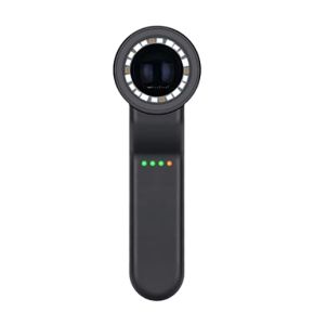
Today, digital dermatoscopes dominate the market. They are equipped with high-resolution cameras that capture detailed images of the skin. These devices allow dermatologists to see microscopic details, enhancing the quality of diagnoses.
Technological innovations include LED lighting, which provides brighter and more consistent light. Polarization is another major advancement. It reduces reflections on the skin, allowing better visualization of underlying structures. These improvements have made the dermatoscope more effective and easier to use.
What Dermoscopy Techniques Are Used by Professionals?
Examination Methods
Dermatologists use different methods to examine the skin with a dermatoscope:
- Contact: The device is placed directly on the skin, often with a gel or oil to improve image clarity. This method is common and allows detailed observation of skin structures.
- Non-Contact: The device is held close to the skin without touching it. This is useful for examining sensitive areas or when direct contact is not recommended, such as in the case of open wounds or infections.
- Immersion: A liquid (like alcohol or oil) is applied to the skin to reduce light reflection. This allows better visualization of internal structures, especially for pigmented lesions.
The choice of method depends on the lesion being examined and the practitioner's preferences. Each technique offers specific advantages for observing different types of lesions.
Interpretation of Dermatoscopic Patterns
The patterns observed during dermoscopy provide valuable information about the nature of skin lesions. Dermatologists look for specific characteristics such as:
- Pigment Networks: They indicate the distribution of melanin in the skin. A regular network suggests a benign lesion, while an irregular network may be suspicious.
- Globules and Dots: Their size, shape, and distribution help differentiate lesion types.
- Radial Lines: They can indicate lesion growth and help identify melanomas.
- Vascular Structures: The shape and arrangement of blood vessels can reveal vascular or inflammatory conditions.
Correct interpretation of these patterns requires specialized training. Dermatologists learn to recognize the characteristic signs of different skin diseases to make accurate diagnoses.
Training for Effective Use
Using a dermatoscope requires specific skills. Professionals undergo training to master examination techniques and image interpretation. This includes:
- Learning the different examination methods and knowing when to use them.
- Understanding dermatoscopic patterns and their clinical significance.
- Staying informed about the latest technological and methodological advancements.
This ongoing training is essential to ensure accurate diagnoses and provide the best care to patients.
What Are the Clinical Applications of the Dermatoscope?
This medical device is a versatile tool used in various clinical situations:
- Detection of Melanoma and Other Skin Cancers: It helps identify suspicious lesions for early intervention. Rapid detection significantly improves treatment success rates.
- Diagnosis of Pigmentary Disorders: Conditions like nevi (moles) or lentigines can be accurately assessed, helping differentiate benign lesions from malignant ones.
- Vascular Conditions: Blood vessel anomalies, such as hemangiomas or rosacea, are easier to diagnose with a dermatoscope.
- Monitoring Skin Lesions: By capturing regular images, dermatologists can track lesion progression over time, detecting subtle changes.
- Evaluation of Inflammatory Diseases: Conditions like psoriasis or eczema display specific features visible with a dermatoscope.
These applications make the dermatoscope an indispensable tool for comprehensive and effective dermatological practice.
How Do New Technologies Improve Dermoscopy?
Digital Dermoscopy
Digitization has transformed dermoscopy, offering new possibilities to professionals:
- Image Storage: Digital dermatoscopes allow for the saving of lesion images. This facilitates patient follow-up and image comparison over time.
- Information Sharing: Images can be shared with other specialists for additional opinions, enhancing collaboration and care quality.
- Medical Documentation: Digital images enrich patient records, providing a visual trace of clinical observations.
Digital dermoscopy also facilitates the use of analysis and diagnostic assistance software.
Computer-Assisted Diagnostic Software
Advances in artificial intelligence have led to the development of software capable of analyzing dermatoscopic images:
- Automatic Analysis: Software can identify specific patterns and alert to suspicious features.
- Increased Accuracy: They help reduce human errors by providing a second opinion.
- Time Savings: Rapid image analysis allows dermatologists to focus on treatment.
While these tools do not replace clinical expertise, they are a valuable complement to improve diagnoses.
Teledermatology
Teledermatology uses technology to provide remote consultations:
- Accessibility: Patients can consult a dermatologist without traveling, which is ideal for rural areas or individuals with limited mobility.
- Speed: Online consultations allow for quicker management of skin conditions.
- Continuous Follow-Up: Patients can regularly send images for effective monitoring.
Teledermatology expands access to specialized care and optimizes the management of medical resources.
Why Choose a Dermatoscope from Placemed?
At Placemed, we are committed to providing high-quality dermatoscopes tailored to professionals' needs:
- Wide Selection: We offer a diverse range of dermatoscopes, from portable models to advanced digital devices.
- Guaranteed Quality: All our products are certified and meet current medical standards.
- Expert Advice: Our team is available to help you choose the model best suited to your practice.
- Competitive Prices: We offer advantageous rates, allowing you to equip your clinic without compromising on quality.
How to Maintain Your Dermatoscope?
To ensure the proper functioning of your dermatoscope, Placemed recommends adhering to certain maintenance guidelines:
- Regular Cleaning: After each use, clean the device with a soft cloth and appropriate products to remove residues and germs.
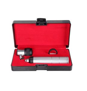
- Proper Storage: Keep the device in its protective case to avoid shocks and dust.
- Component Checks: Regularly inspect the condition of lenses, light sources, and batteries to ensure optimal performance.
- Software Updates: For digital models, ensure that software is updated to benefit from the latest features and improvements.
Proper maintenance guarantees the reliability of your device and the quality of your dermatoscopic examinations.
The dermatoscope is an essential tool for dermatologists and skin health professionals. It enhances diagnostic accuracy, enables early disease detection, and contributes to higher quality patient care. With technological advancements, this device continues to evolve, offering increasingly advanced features.
At Placemed, we are proud to offer a selection of dermatoscopes that meet the varied needs of practitioners. Our commitment to quality and service makes us the ideal partner to equip your clinic. Explore our range today and discover how a quality dermatoscope can transform your practice.
 Francais